What is MRI and How Does it Work?
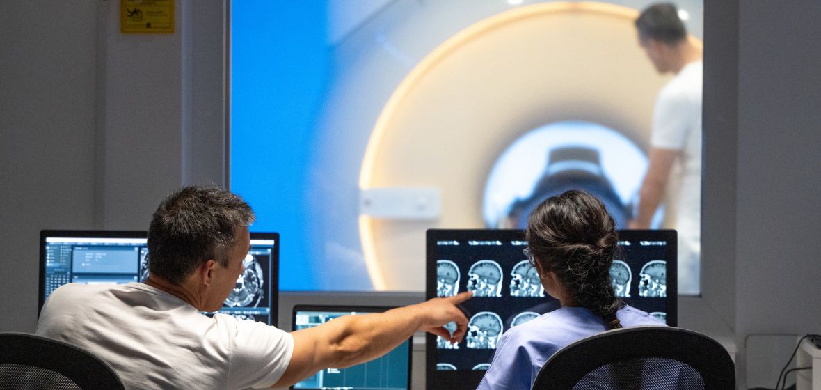
Magnetic resonance imaging (MRI) is a medical imaging technique used to visualize detailed internal structures without the use of x-rays. An MRI scanner consists of a large and very strong magnet in which the patient lies down. A radio wave is used to send signals to the body and then receive signals back. These returning signals are converted into pictures by a computer attached to the scanner. Pictures of almost any part of your body can be obtained at almost any angle.
Our facility offers the most advanced MRI techniques and procedures not offered at many MRI centers across the country. This requires a high level of training and knowledge offered by our nationally registered MRI technologists.
What are the uses and advantages of an MRI scan over other types of scans?
- MRI is excellent at looking at the non-bony parts or “soft tissues” of the body. In particular, the brain, spinal cord and nerves are seen much more clearly with MRI than with regular x-rays and CAT scans.
- Muscles, ligaments and tendons are seen quite well so that MRI scans are commonly used to look at knees and shoulders following injuries.
- All UofL Hospital MRI scanners have large bore openings and are open on both ends reducing anxiety (claustrophobia).
- Effects of strokes can be seen within minutes. Brain function and activity can be seen instantly with fMRI (functional MRI) techniques.
- 3D imaging can be acquired to assist surgeons by special computers in the operating room providing a “road map” to avoid vital areas.
- MRI scanner uses no x-rays (radiation) and poses no known biological hazards.
IMPORTANT: Patients with pacemakers, certain aneurysm clips, cochlear ear implants, or mechanically activated devices, could be contraindications to enter the MRI room. However, many metallic implants CAN be imaged. If you have received an ID card for any implant, please give it to the technologist to verify its safety in an MRI environment.
What will you experience during your MRI scan?
Upon Arrival
- The technologist will interview you to obtain a medical history and metal screening.
- The technologist will explain your exam and answer any questions.
- You will be asked to change into a gown.
- A member of our staff will take you to the MRI room, help you onto the table and position you for the scan. You will be given ear plugs, or in some cases headphones, to reduce the noise associated with taking the MRI pictures.
During your MRI scan
- While taking pictures, you will hear a loud “knocking” noise. This is normal.
- It is VERY important that you do not move any part of your body. The slightest movement will cause your images to blur, in many cases, requiring repeating or resulting in a limited diagnostic report.
- Your exam is composed of multiple series of scans. Each of these scans takes approximately three to five minutes each. Typically, a routine exam consists of five to seven series for a total of about 30-45 minutes.
- Some exams allow you to listen to music while having your MRI, others are given ear plugs. In either case, you will be able to communicate with the technologist during your scan through an embedded microphone within the MRI scanner.
If you have questions about imaging, contact UofL Health – Diagnostic Imaging & Radiology at 502-562-3257.