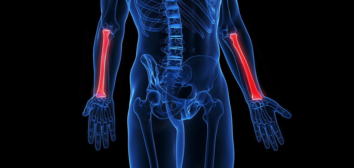
A distal radius fracture occurs at the lower end of the radius bone in the forearm, just above the wrist. It is by far the most common fracture that occurs in humans, accounting for nearly 20% of all bone breaks.
Falling on an outstretched hand accounts for most of these injuries. The reason is simple.
If you fall, it is natural to extend your hands to break the impact of the fall and thus protect more important parts of the body like the head and chest. Doing so automatically transfers the full force of the fall to the hand-wrist area and thus leads to a fracture (or break) of the distal radius.
Distal radius fractures are common across all age groups.
Children are simply very active and prone to falls while at play. In this age group, fractures may often involve the “growing end” of the distal radius. Such injuries are called “growth plate injuries” and may on occasion affect the future growth of the bone.
Fractures in older adults occur because of a combination of two factors: older adults are prone to falling because of loss of body strength or lack of balance, and they are more likely to have osteoporosis, making it easier for bones to break.
Diagnosis
Distal radius fractures may vary in degrees of severity. At its simplest, the break may merely be a “crack” in the bone without any displacement of the bone pieces. At its most complex, the distal radius may shatter into multiple pieces involving the joint (articular surface) and may even be accompanied by dislocation of the wrist joint.
X-ray images are the best way to diagnose distal radius fractures. Typically, at least two pictures of the wrist area are taken, each picture being at right angles to the other. The first picture, called the postero-anterior or PA view, provides a front-to-back image of the bones while the second, called the lateral view provides a side-to-side image.
These two images help a hand surgeon assess the amount of shattering as well as the degree the bones are displaced.
Sometimes, additional X-ray views may be needed and on occasion, more advanced imaging methods like MRI or CT scans can also become necessary. These additional investigations enable the surgeon to better assess the degree the bones are displaced and look for subtle breaks, associated ligament injuries and other soft tissue injuries that can accompany distal radius fractures.
A break in the forearm bone beside the radius, known as the ulna, is a very common injury that accompanies distal radius fractures. Most often, only the tip of the ulna, known as the ulnar styloid, is fractured.
The ulnar styloid is attached to an important ligament complex known as the TFCC (Triangular Fibro Cartilage Complex). This is an important ligament for stabilizing the joint between the distal radius and ulna, called the DRUJ (Distal Radio Ulnar Joint).
UofL Health – Kleinert Kutz Hand Care is at the forefront of pioneering treatments and comprehensive care for various hand and upper extremity conditions. Our dedicated team of specialists comprises world-renowned hand surgeons, therapists and support staff, all committed to delivering the highest level of care to our patients. If you are seeking specialized care for your hand or upper extremity condition, do not hesitate to schedule an appointment with Kleinert Kutz Hand Care by calling 502-561-4263.